Cathodoluminescence
microscopy
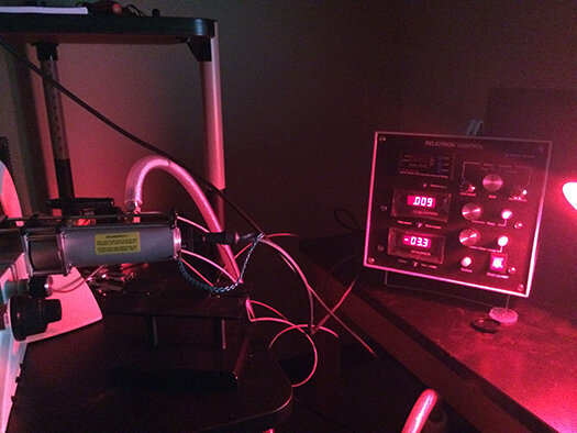
A Relion Reliontron cold cathodoluminescence is mounted on a Leica DM2700 P. Luminescence is usually triggered and quenched by manganese and iron content, respectively. Cathodoluminescence thus allows the characterization of cement phases and relate their chemistry to the fluid from which their precipitated, especially when combined with punctual carbon and oxygen stable isotope composition measurement.
The facility is accessible after taking a UTSA mandatory online course on Lab safety and X-ray safety.
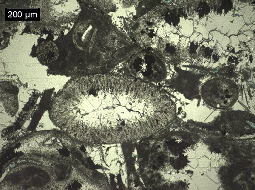
Skeletal grainstone in the Corbula Bed of the Glen Rose Formation (Albian, Early Cretaceous), Boerne, Texas, plane polarized light.
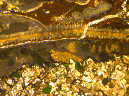
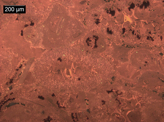
Skeletal packstone (limestone) with phosphatic matrix, bivalves allochems, glauconite, dolomite and quartz crystals. Altmann Member (late Hauterivian-early Barremian, Early Cretaceous), Pilatus, Switzerland, plane polarized light.
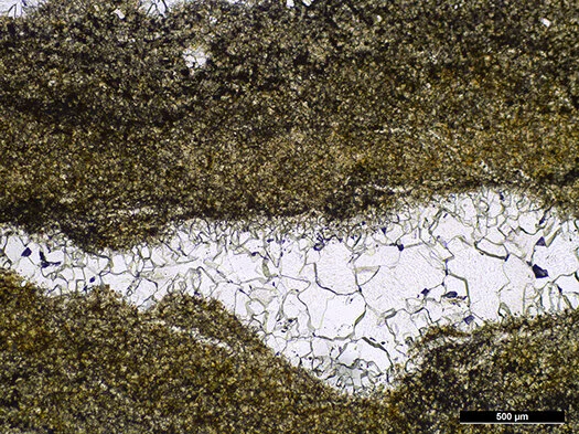
Birdseye in stromatolitic limestone, Glen Rose Formation (Albian, Early Cretaceous), Boerne, Texas, plane polarized light.
Skeletal grainstone in the Corbula Bed of the Glen Rose Formation (Albian, Early Cretaceous), Boerne, Texas, cathodoluminescent light.
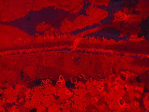
Skeletal packstone (limestone) with phosphatic matrix, bivalves allochems, glauconite, dolomite and quartz crystals. Altmann Member (late Hauterivian-early Barremian, Early Cretaceous), Pilatus, Switzerland, cathodoluminescent light.
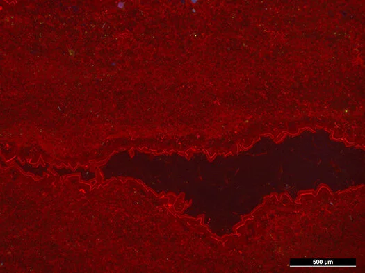
Birdseye in stromatolitic limestone, Glen Rose Formation (Albian, Early Cretaceous), Boerne, Texas, cathodoliminescence light.
Wavelength Dispersive X-ray fluorescence spectrometry (WD-XRF)
A Rigaku Primus II WD-XRF permits the chemical analysis of fused, powdered and liquid samples, from Boron to Uranium.
Sample preparation includes sawing and powdering, preparation of fused beads with Lithium Tetraborate using a Claisse LeNeo fluxer, and calculation of the Loss On Ignation (LOI) using a muffle oven. We recently acquired a hydraulic press and a die set to produce pressed powder beads that will improve detection limits for trace elements.
Fused beads are subsequently analyzed with the WD-XRF, using an easy scan, qual or quant method.
The lab is accessible after taking a UTSA mandatory online course on Lab safety and X-ray safety.
Contact me for training, schedule and pricing.